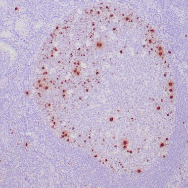
Phosphohistone H3 (PHH3) is a marker specific for cells undergoing mitosis. Serine 10 of Histone H3 is phosphorylated in association with mitotic chromatin condensation in late G2 and M phase of the cell cycle and thus, PHH3 can distinguish mitosis from apoptotic nuclei. The range of percentage PHH3 positive tumor nuclei is from 0.0 to 6.6% (median value 0.8%). Increased expression of PHH3was significantly associated with tumor thickness (p = 0.031), presence of tumor ulceration (p =0.041) and tumor necrosis (p =0.027), but not with Clark’s level of invasion. High levels of PHH3 is associated with increased mitotic count (p = 0.003) and high Ki-67 expression (p = 0.002). For central nervous system tumors, melanoma, soft tissue tumors, GIST, etc., PHH3 mAb is helpful for tumor pathological classification and prognosis.

