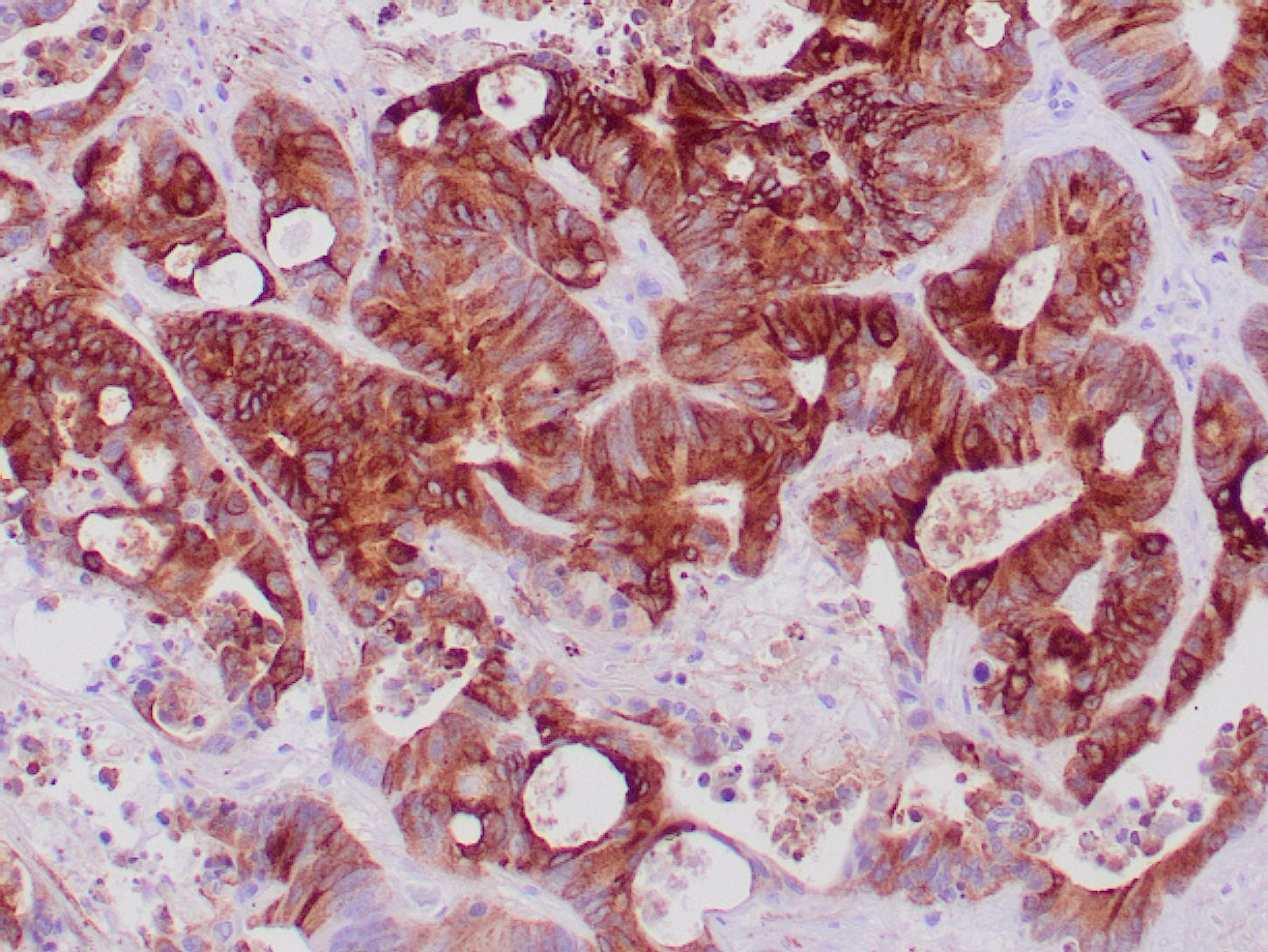

In Western blotting, it recognizes proteins in the MW range of 265-400kDa, identified as different glycoforms of EMA. EMA may provide a protective layer on epithelial cells against bacterial and enzyme attacks. In immunohistochemical assays, it superbly stains routine formalin/paraffin carcinomas. Anti-EMA antibody is a valuable marker for staining many carcinomas. It stains normal and neoplastic cells from various tissues, including mammary epithelium, sweat glands, and colorectal carcinoma. Hepatocellular carcinoma, adrenal carcinoma, and embryonal carcinomas are consistently EMA negative, so keratin positivity with negative EMA favors one of these tumors. EMA is frequently positive in meningioma, which can be helpful when distinguishing it from other intracranial neoplasms such as schwannomas. Antibody to EMA is helpful as a pan-epithelial marker for detecting early metastatic loci of carcinoma in bone marrow or liver.